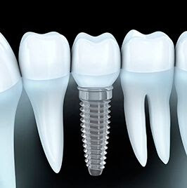
what are dental xrays
DO YOU WANT TO KNOW ABOUT THE X- RAYS TAKEN BY YOUR DENTIST
x rays in dentistry shows clearly the in and around anatomical structures of teeth ,which can not be seen over clinical examination.It is an easy way also.This helps dentist to diagonse the problems and treatment plan in an very precise manner.
There are many types of dental xrays in the field.Lets know all about it,in one by one.
1.PERIAPICAL INTRA ORAL XRAY
This type of xrays is the most commonly used one in dental field.This will show the exact structures of not only teeth,but also the surrounding structures like gums,tissues,blood vessels and bone.But this is not a three dimensional x ray.This is only two dimensional x-ray.First your dentist will place a dental film inside your mouth in paticular region,this x ray covers maximum of four teeth.The most important thing here is,you should not change your position when your dentist takes your x-ray,then a cone shaped x ray machine will be placed outside the region where the film is kept inside. You will hear a beep sound,indicates that the x-ray is taken.The mechanism behind this x-ray is teeth absorbs more rays than the gums and tissues,so teeth appears brighter where the decay /infections looks darker as they receives less rays from the exposure.
2.RADIO VISIO GRAPHY (RVG)
This is also the same mechanism that the periapical intra oral x ray has.the major difference between them is,in RVG x ray films are not used,instead x ray sensor is used.This is connected to system,so once the beep sound is heard ,the x ray image will be automatically displayed on the screen within a second,where in periapical intra oral radiograph the x ray films are to be developed.This is mostly taken during root canal treatment.
3.DENTAL PANORAMIC RADIOGRAPH(OPG)
OPG ,this is also called as full mouth x ray commonly to the patients.This will covers all the teeth in mouth and also the other external parts like sinus,nerves,maxilla and mandible,orbital and nasal.This gives dentist the exact bone quantity,quality,alignment of the teeth.During opg ,patient should remove gold or other ornaments.like chain,nose piercing,ear rings,because their images will also reflect in this opg. Patient is asked to wear lead sheet apron over them during their exposure time,to protect them from radiation.This is also two dimensional x ray & mostly used for impacted tooth,implants,gingivitis.
4.CONE BEAM COMPUTED TOMOGRAPHY(CBCT)
The first three dimensional x ray in dentistry.It shows the structures of teeth in three dimensional view. very useful for surgery cases.This is similar to MRI scans,which most of us are known well.This will recommend by the dentist for very rare cases.
We in MSR dentistry has all the x rays mentioned above.
MSR DENTISTRY AND IMPLANT CENTER.
Address:-
Vallarlar Illam NO 68,
Anna Street,
Chitlapakkam,
Chennai - 600064.
Landmark : Near Gangai amman koil
CONTACT :044-42142777


Comments
Post a Comment